


Advances in technology and improvement in modern health infrastructure have led to exciting developments in dentistry imaging. 3D Dentistry is one of the most powerful and revolutionary tools that has improved precision in diagnosis and led to more effective treatments and overall better patient outcomes.
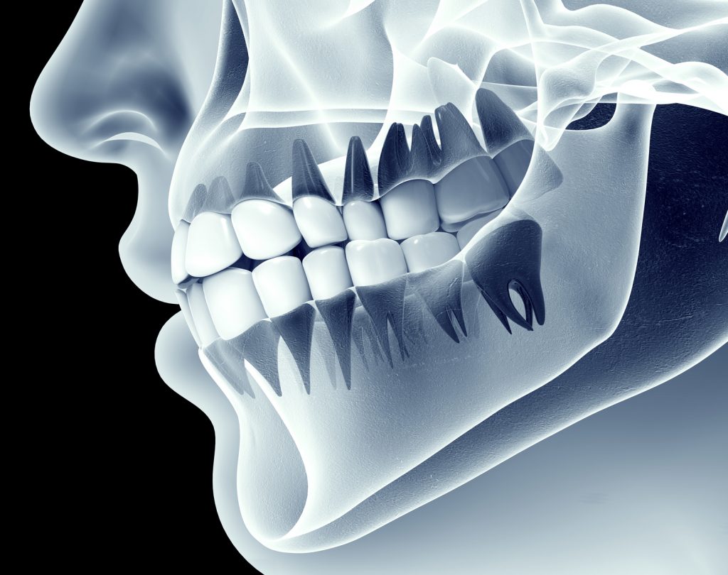
3D Dentistry allows dentists to see accurate, detailed three-dimensional images of a patient’s mouth, teeth, and skull and you can track every information about 3D digital modeling of your patients dental condition with our orthodontic practice management software.
Unlike conventional 2D x-rays, 3D X-rays are designed to help a dentist to understand the entire mouth and examine it in slices similar to a CT scan. They do this by capturing the real 3-dimensional image of the mouth, teeth, bones, soft tissues, nerve pathways, etc.
3D Dentistry in details
The medical imaging technique involved in 3D Dental Imagery is called Cone beam computed tomography (CBCT). In CBCT, the x-rays divergent or opposing, which leads to the formation of a cone-beam shaped beam. The use of a cone-shaped beam enables dentists to use the information they need without much radiation to patients.
CBCT machines are now commonplace among technologically up-to-date dental clinics across the world. They are vital contributors to quality dental care that help dentists to formulate effective approaches better to treat dental conditions.
3D dental technology also displays things in extensive detail which provide for better patient education. Patients can visualize and clearly comprehend their dental x-rays, which enhances their experience.
3D dental images can be used for; aiding surgical planning for impacted teeth, accurate placement of implants, reconstructive surgery, evaluating bone structure and tooth orientation, detection, assessment and diagnosis of jaw tumors, reconstructive surgery, identifying the origin of pain and diagnosis of Temporomandibular joint disorder (TMJ).
With 3D technology, dentists can plan implants for their patients in a 3 dimensional way. There is no chance for guesswork. You will know the exact thickness, heights, volume, how much bone you have. Use of cone beam technology also enables a dentist to safely identify nerve or arteries that could lead to surgical complications.
Previously, the promising technology has, however, made little progress outside maxillofacial surgery and implantology, where 3D dental imagery is an absolute must. The resulting DICOM (Digital Imaging and Communications in Medicine) images, which were generally large, complex, and challenging to manage, especially those stored in a cloud-hosted imaging system. Early adopters embraced CAD/CAM milling by Cerec, Sinterex, and others to create crowns, bridges, inlays.
Now, most dentists and facility managers agree that digital 3D impressions fabrications will replace traditional methods of creating crowns, bridges, inlays, dental bars, etc.
Digital 3D printing is much faster, easier, and more accurate than CAD/CAM milling. Also, CAD/CAM milling uses ceramics such as porcelain CEREC® Blocs while Digital 3D printing uses resins, adding more layers until it’s complete. Newer models can melt non-precocious metal alloys in powder form and layer them using 3D printing.
Digital 3D printing may be pricey, but technology-minded dentists who have tried these machines say it’s worth the investment. This is especially true, considering that the equipment will reduce your total procedure time will be reduced from 2-3 appointments to a single one.
The accuracy that comes with digital 3D printing is a definite advantage that provides better diagnosis, better treatment protocols specific to the patients’ unique dental needs, and, therefore, increased patient satisfaction. Furthermore, these equipment use fewer materials, which increases revenues and profits.
Another key benefit of 3D Dentistry is speed and convenience. A 3D X-ray takes only about 14-20 seconds with powerful computers finishing construction in just 7 minutes instead of 50 minutes.
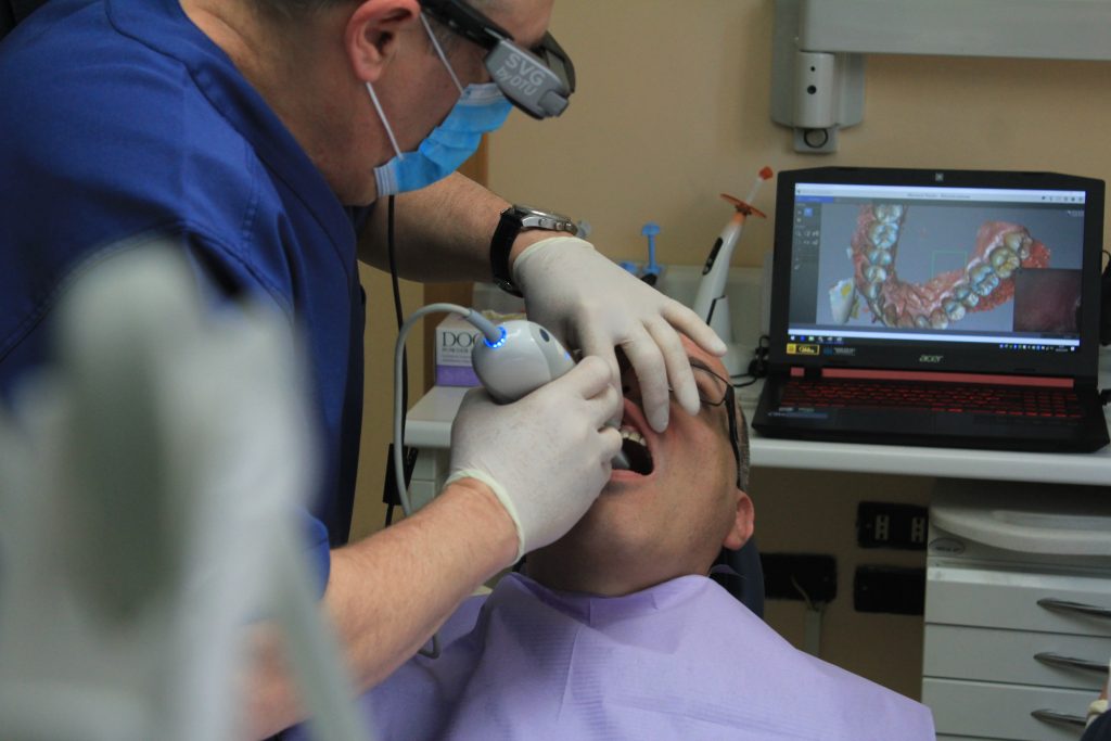
To fulfill the growing demand for safe, highly accurate and innovative digital optical scanners by dentistry professionals, more and more companies are releasing new models into the market with unique features such as 3Shape’s Trios portable wireless scanner or the more affordable Densys3D.
What’s more, users can run most of these newer models from a standard high-speed laptop that fits well into smaller dental offices, unlike conventional first-generation cart-placed scanners like iTero.
These new models can run in a variety of applications for ease of use and convenience. 3D viewing and dental CAD-CAM applications support optical scanners that can export directly to STL format.
STL files are quite versatile in newer models; you can create them on one machine, edit them on another, and even send them to a third machine in a different laboratory for final printing. It is also possible to print all 3D models exported in an STL format fairly cheaply in the office through 3D printers such as FormLabs’ Form2.
Affordable 3D scanners such as AutoScan by Shining3D can convert dental stone models into 3D digital STL models for digital viewing.
Final Thoughts
3D Dentistry is clearly the new standard of dental care due to its accurate diagnosis, better results, efficient treatment protocols, and improved patient satisfaction. Technology minded dentists, facility managers, researchers, and laboratories agree that there is no ceiling to the capabilities of 3D dental imagery in the future.